Fig 3. - Median Sections through normal and Down's Syndrome Skulls.
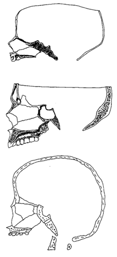 Fig 3.gif (26730 bytes)
|
Fig 3
Normal foetal skull (redrawn after Bosma[4])
Normal adult skull (redrawn after Gray[54])
Down's Syndrome skull (redrawn after Benda[2]) |
MEDIAN SECTIONS THROUGH NORMAL and DOWNS SYNDROME SKULLS
At birth the spheno-basilar synchondrosis is patent and the pre and post sphenoid are unfused. Note that the occiput and sphenoid are in a near straight line and at an angle of about 45o to the horizontal.
- In the adult observe the large size of the sphenoid sinus and frontal sinuses and note their influence on the configuration of the skull and face. The cranio-facial angle, measured at the midpoint of the optic chiasmal groove from the lines intersecting from nasion and basion, represents the degree of cranial flexure and normally should have a value of about 130o.
- In Downs syndrome the sphenoid sinus is missing and the sphenoid body is hypoplastic. The absence of the frontal sinus results in a straight, infantile forehead. The cranial base is still nearly in a straight line, as in the foetal skull, but the angle to the horizontal is steeper. The anterior cavity of the cranium is shorter, steeper and on a higher plane than normal. There is absence of diploe in the vault bones, resulting in a thin, light skull. Nasion is retracted and the maxilla is hypoplastic.